Brain Tumor Segmentation using Convolutional Neural Networks in MRI Images
Sérgio Pereira, Adriano Pinto, Victor Alves and Carlos A. Silva
University of Minho
Accepted at IEEE TMI
Abstract
Among brain tumors, gliomas are the most common and aggressive, leading to a very short life expectancy in their highest grade. Thus, treatment planning is a key stage to improve the quality of life of oncological patients. Magnetic Resonance Imaging (MRI) is a widely used imaging technique to assess these tumors, but the large amount of data produced by MRI prevents manual segmentation in a reasonable time, limiting the use of precise quantitative measurements in the clinical practice. So, automatic and reliable segmentation methods are required; however, the large spatial and structural variability among brain tumors make automatic segmentation a challenging problem. In this paper, we propose an automatic segmentation method based on Convolutional Neural Networks (CNN), exploring small 3×3 kernels. The use of small kernels allows designing a deeper architecture, besides having a positive effect against overfitting, given the fewer number of weights in the network. We also investigated the use of intensity normalization as a preprocessing step, which though not common in CNN-based segmentation methods, proved together with data augmentation to be very effective for brain tumor segmentation in MRI images. Our proposal was validated in the Brain Tumor Segmentation Challenge 2013 database (BRATS 2013), obtaining simultaneously the first position for the whole, core and enhancing regions with Dice Similarity Coefficient (DSC) metric (0.88, 0.83, 0.77) for the Challenge data set. Also, it obtained the overall first position by the online evaluation platform.
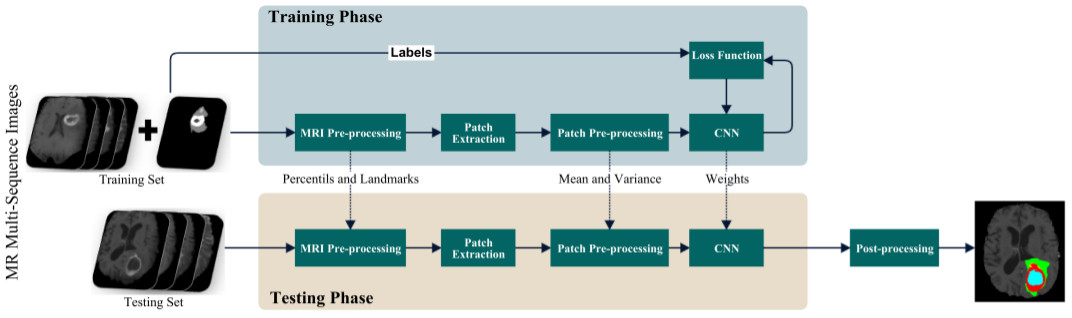
Proposed Pipeline
Some Segmentations

Supplementary Material
Additional material: CNN weights, other figures and tables with more experiments not described in the article for lack of space.
If you use the weights or any result for your study, please cite: S. Pereira, A. Pinto, V. Alves and C. A. Silva, "Brain Tumor Segmentation Using Convolutional Neural Networks in MRI Images," in IEEE Transactions on Medical Imaging, vol. 35, no. 5, pp. 1240-1251, May 2016.
Acknowledgements
This research work was supported by the Center for Microelectromechanical Systems – MEMS-UMinho Research Unit. Sérgio Pereira was supported by a scholarship from the Fundação para a Ciência e Tecnologia (FCT), Portugal (scholarship number PD/BD/105803/2014). Brain tumor image data used in this article were obtained from the MICCAI 2013 Challenge on Multimodal Brain Tumor Segmentation. The challenge database contain fully anonymized images from the Cancer Imaging Atlas Archive and the BRATS 2012 challenge.